How to Become an EKG Technician: The Best Books, In Order
This curriculum takes a complete beginner from zero knowledge of the heart all the way through EKG/ECG certification readiness. Each stage builds directly on the last: you first learn the anatomy and physiology that makes the heart tick, then decode basic rhythms, then master the clinical 12-lead interpretation used on the job, and finally drill the exam-style questions needed to pass your certification. Expect roughly 8–10 books total, chosen for their widespread use in allied health programs and EKG technician training courses.
Heart Anatomy & Physiology Foundations
BeginnerUnderstand the structure of the heart, the cardiac conduction system, and how electrical impulses produce a heartbeat — the essential vocabulary for everything that follows.
▸ Study plan for this stage
Pace: 4–5 weeks, ~25–30 pages/day. Start with Thibodeau's heart anatomy and conduction chapters (approximately 40–50 pages), then move to Moini's cardiac physiology sections (approximately 30–40 pages). Include 2–3 review days per week.
- The four chambers of the heart and their roles in blood flow (atria vs. ventricles)
- The coronary circulation system and how the heart receives its own blood supply
- The cardiac conduction system: SA node, AV node, bundle of His, Purkinje fibers, and the sequence of electrical activation
- The relationship between electrical impulses and mechanical contraction (excitation-contraction coupling)
- The cardiac action potential and ion movement (sodium, potassium, calcium) across the cell membrane
- Normal sinus rhythm and the intrinsic heart rate
- Anatomical landmarks on the chest wall for lead placement and understanding cardiac vectors
- What are the four chambers of the heart, and what is the function of each?
- Trace the path of electrical conduction through the heart, naming each structure in sequence and the approximate time it takes.
- How does the SA node function as the heart's natural pacemaker, and what happens if it fails?
- What is the relationship between the cardiac action potential and the mechanical heartbeat?
- How does coronary circulation differ from systemic circulation, and why is it critical for heart function?
- What anatomical landmarks on the chest wall correspond to the standard EKG lead positions?
- Create a labeled diagram of the heart showing all four chambers, valves, major vessels, and the conduction pathway; color-code the conduction system.
- Trace the electrical impulse through the heart on a blank diagram 5 times without reference, timing yourself to build automaticity.
- Using Thibodeau's illustrations, identify the SA node, AV node, bundle of His, and Purkinje fibers on progressively unlabeled diagrams.
- Explain out loud (or write) the sequence of atrial depolarization, ventricular depolarization, and repolarization as if teaching someone else.
- Locate the anatomical landmarks for standard EKG lead placement (V1–V6, I, II, III, aVR, aVL, aVF) on a torso model or diagram.
- Create a comparison table: normal conduction times at each node/structure (SA node delay, AV node delay, ventricular conduction time) and what happens clinically when each is prolonged.
Next up: This foundation in cardiac anatomy and the normal electrical conduction pathway enables you to recognize deviations from normal on an EKG—arrhythmias, blocks, and ischemia all represent disruptions to this baseline electrical and mechanical process.

A classic allied-health anatomy and physiology text written for beginners; its dedicated cardiovascular chapters give you the structural and functional vocabulary you need before touching an EKG machine.

Bridges general A&P directly to clinical health careers, reinforcing cardiac conduction concepts in plain language — ideal as a second pass before moving into rhythm interpretation.
Introduction to EKG Basics & Rhythm Recognition
BeginnerRead and measure the components of an EKG strip (P wave, QRS, T wave, intervals), identify normal sinus rhythm, and recognize the most common arrhythmias with confidence.
▸ Study plan for this stage
Pace: 8–10 weeks, ~25–35 pages/day. Start with Atwood (2–3 weeks), move to Ellis (2–3 weeks), then Dubin (3–4 weeks) to build from foundational concepts to rapid interpretation skills.
- Cardiac action potential and the electrical conduction system (SA node, AV node, His bundle, Purkinje fibers) as covered in Atwood
- EKG paper mechanics: time and voltage measurements, grid interpretation, and how to calculate heart rate using the 6-second strip method (Ellis)
- Normal waveform anatomy: P wave, QRS complex, T wave, U wave, and their clinical significance (Atwood and Ellis)
- Intervals and segments: PR interval, QRS duration, QT interval, and ST segment—normal values and what deviations mean (Ellis and Dubin)
- Normal sinus rhythm criteria and how to systematically analyze any rhythm strip (Ellis and Dubin)
- Recognition of the most common arrhythmias: sinus tachycardia, sinus bradycardia, atrial fibrillation, atrial flutter, ventricular tachycardia, and premature contractions (Atwood, Ellis, and Dubin)
- Systematic rhythm strip analysis: rate, regularity, P waves, PR interval, QRS duration (Dubin's methodical approach)
- Clinical correlation: understanding why arrhythmias occur and their hemodynamic significance (Atwood and Ellis)
- What are the normal values for the PR interval, QRS duration, and QT interval, and what do prolongations or shortening of each indicate?
- How do you calculate heart rate from an EKG strip using at least two different methods (6-second strip and R-R interval methods)?
- What are the five criteria for normal sinus rhythm, and how would you systematically evaluate a rhythm strip to confirm it?
- Describe the characteristic features of atrial fibrillation, atrial flutter, and ventricular tachycardia—how would you distinguish between them on a strip?
- What is the relationship between the cardiac action potential and the EKG waveforms (P wave, QRS, T wave)?
- Given a rhythm strip with premature contractions, how would you determine whether they are premature atrial contractions (PACs) or premature ventricular contractions (PVCs), and what is the clinical significance of each?
- Using Atwood's diagrams of the conduction system, trace the electrical pathway from the SA node through the ventricles and correlate each segment to the corresponding EKG waveform.
- Practice measuring EKG paper: mark 10 consecutive small squares and 5 consecutive large squares on graph paper, then use Ellis's grid method to measure intervals on 20 different rhythm strips (normal and abnormal).
- Calculate heart rate using both the 6-second method and the R-R interval method on 15 strips from Ellis and Dubin; compare results to verify accuracy.
- Analyze 30 rhythm strips (10 normal sinus, 10 with common arrhythmias, 10 mixed) using Dubin's systematic approach: rate → regularity → P waves → PR interval → QRS duration. Document findings for each.
- Create flashcards for the 6–8 most common arrhythmias (sinus tach/brady, AFib, flutter, VT, PACs, PVCs) with characteristic features, normal values, and clinical significance from Ellis and Dubin.
- Perform a "blind" rhythm strip challenge: have a peer or instructor present 10 unlabeled strips; identify the rhythm and explain your reasoning using the systematic method from Dubin.
Next up: Mastery of basic rhythm recognition and waveform measurement establishes the foundation for the next stage, which will deepen your ability to recognize complex arrhythmias, interpret 12-lead EKGs, and correlate findings with clinical symptoms and treatment decisions.
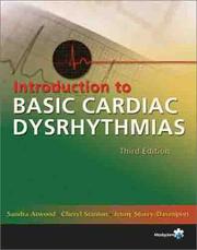
One of the most widely adopted entry-level EKG texts in allied health programs; it walks through each waveform component step-by-step with abundant practice strips, making it the perfect first rhythm book.
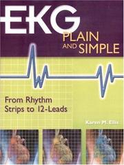
Builds directly on Atwood's foundation by adding more rhythm examples and a systematic strip-analysis method, reinforcing pattern recognition before you advance to 12-lead work.
A legendary self-teaching classic used by millions; its programmed, step-by-step format cements rhythm logic and waveform meaning in a memorable, visual way — read it here to solidify your beginner knowledge before going deeper.
12-Lead EKG Interpretation
IntermediateUnderstand the 12-lead EKG system, lead placement, axis determination, bundle branch blocks, ST changes, and myocardial infarction patterns used daily by EKG technicians and clinical staff.
▸ Study plan for this stage
Pace: 8–10 weeks, ~25–35 pages/day with 2–3 days/week dedicated to practice strips and axis calculations
- The 12-lead EKG system: understanding the 6 limb leads (I, II, III, aVR, aVL, aVF) and 6 precordial leads (V1–V6) and what each lead visualizes
- Proper electrode placement and lead orientation to ensure accurate recordings and avoid misinterpretation
- Mean QRS axis determination using the quadrant method and hexaxial reference system
- Bundle branch blocks (RBBB and LBBB): recognition criteria, QRS duration, and characteristic patterns in specific leads
- ST-segment changes: elevation, depression, and T-wave inversions as indicators of ischemia and injury
- Acute myocardial infarction (AMI) patterns: identifying location (anterior, inferior, lateral, posterior) based on lead involvement and reciprocal changes
- Systematic interpretation approach: rhythm, rate, axis, intervals, segments, and clinical correlation
- Common EKG artifacts and mimics: distinguishing pathology from technical error or normal variants
- How do you systematically approach a 12-lead EKG interpretation, and what are the key parameters you assess in order?
- Given a 12-lead EKG, can you identify which coronary artery is likely occluded based on ST elevation location and reciprocal changes?
- What are the diagnostic criteria for RBBB and LBBB, and how do they differ in QRS morphology across the precordial leads?
- How do you calculate the mean QRS axis using the quadrant method, and what axis ranges indicate normal, left axis deviation, and right axis deviation?
- What is the clinical significance of ST-segment elevation versus depression, and how do you differentiate acute MI from other causes of ST changes?
- How does proper electrode placement affect EKG interpretation, and what artifacts can result from improper placement or patient movement?
- Complete 50+ practice 12-lead EKG strips from Thaler's book, focusing on rhythm identification and basic measurements before moving to pathology
- Use the hexaxial reference system to manually calculate the mean QRS axis on 20 different EKGs; compare your results with provided answers
- Create a visual reference chart mapping each of the 12 leads to the anatomical area of the heart they visualize, and identify which coronary arteries supply each region
- Interpret 30 EKGs from Green's book that demonstrate bundle branch blocks, categorizing each as RBBB or LBBB and explaining the diagnostic criteria you used
- Work through 25 acute MI case studies from Springhouse, identifying the infarction location, culprit artery, and reciprocal lead changes for each
- Perform a mock EKG recording on a peer or mannequin, ensuring correct electrode placement for all 12 leads, then compare your recording with a reference standard
Next up: Mastery of 12-lead interpretation and pattern recognition establishes the foundation for the next stage, which will focus on advanced pathology (arrhythmias, electrolyte abnormalities, and special populations) and clinical decision-making in real-world EKG technician scenarios.
A highly readable, humor-infused intermediate text that systematically teaches 12-lead concepts — axis, hypertrophy, ischemia, and infarction — making it the natural bridge from rhythm strips to full 12-lead reading.

Specifically designed for technicians and nurses learning 12-lead interpretation; its workbook-style practice reinforces lead placement and pattern identification in a hands-on format.

Part of the trusted 'Incredibly Easy' allied-health series, this book consolidates 12-lead concepts with visual mnemonics and clinical context, serving as an excellent review and reference before exam prep.
Certification Exam Preparation
IntermediateApply all prior knowledge under timed, exam-style conditions; master the content domains tested by the CET (Certified EKG Technician) and NHA EKG exams and walk into test day with confidence.
Mometrix's exam-secrets format breaks down every tested domain with concise review notes and test-taking strategies, making it the ideal final review to maximize your score on certification day.
Discussion
Keep reading
Paths that share books, cover the same subject, or open a related topic.