How to Become a Nuclear Medicine Technologist: The Best Books, In Order
This curriculum is designed for learners who already have a foundational science background and want to build the specialized knowledge required to work as a Nuclear Medicine Technologist — from imaging physics and instrumentation, through radiopharmaceuticals and patient procedures, to board examination readiness and licensure. Each stage builds directly on the last: instrumentation vocabulary unlocks radiopharmaceutical logic, which in turn makes board-prep review far more productive.
Imaging Physics & Instrumentation
IntermediateUnderstand how nuclear medicine imaging systems work — from radiation physics and detector design to SPECT and PET instrumentation — giving you the technical language needed for every later stage.
▸ Study plan for this stage
Pace: 8–10 weeks, ~40–50 pages/day (mix of dense physics chapters and technical reference sections)
- Radioactive decay modes (alpha, beta, gamma) and how they relate to nuclear medicine isotope selection
- Interaction of radiation with matter: photoelectric effect, Compton scattering, and pair production in detector materials
- Scintillation detector design and operation: crystal composition, photomultiplier tubes (PMTs), and light collection efficiency
- Gamma camera (Anger camera) architecture: collimators, crystal, PMT array, electronics, and image formation
- SPECT instrumentation: multi-detector systems, gantry mechanics, rotation protocols, and 3D reconstruction principles
- PET instrumentation: coincidence detection, annihilation radiation physics, detector block design, and timing resolution
- Energy resolution, spatial resolution, and sensitivity as performance metrics for imaging systems
- Quality control and calibration procedures for maintaining detector and imaging system performance
- Explain the physical basis for why Tc-99m and F-18 are preferred isotopes in nuclear medicine, referencing their decay characteristics and detector interaction properties
- Describe the complete signal pathway in a gamma camera from photon detection through PMT output to final image formation
- Compare and contrast SPECT and PET instrumentation in terms of detector design, coincidence requirements, and spatial resolution capabilities
- What is the role of collimation in SPECT imaging, and how does collimator design affect sensitivity and spatial resolution trade-offs?
- Explain how Compton scattering affects image quality in both SPECT and PET, and what correction methods are used
- Describe the components and function of a scintillation detector, including how light is converted to electrical signal
- Work through Cherry's decay mode calculations: determine the energy and particle types for 5 common nuclear medicine isotopes, then predict which will interact best with a NaI(Tl) crystal
- Create a labeled diagram of a gamma camera (Anger camera) and trace the signal pathway from incoming photon to digital image, annotating each component's function
- Compare detector specifications (energy resolution, spatial resolution, sensitivity) for at least 3 different gamma camera systems; explain trade-offs using physics principles from Cherry
- Solve 4–5 problems on photon interaction cross-sections and attenuation in tissue using Cherry's chapters on radiation interactions; calculate depth-dependent count loss
- Build or sketch a simple SPECT acquisition protocol: specify collimator type, number of projections, rotation angle, and energy window, justifying each choice with physics reasoning
- Perform a quality control exercise: interpret flood field images and energy spectra from a gamma camera; identify artifacts and propose corrections based on detector physics
Next up: Mastery of imaging physics and instrumentation provides the technical foundation to understand image acquisition protocols, reconstruction algorithms, and clinical optimization strategies in the next stage.

The definitive, widely adopted text on nuclear medicine physics. Read this first to build rigorous understanding of radioactive decay, detector physics, SPECT, and PET before tackling clinical applications.
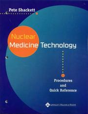
Bridges pure physics to practical imaging procedures. Read after Cherry to see how instrumentation principles translate into real-world technologist workflows and quality control protocols.
Radiopharmaceuticals & Radiation Biology
IntermediateUnderstand how radiopharmaceuticals are produced, labeled, and behave in the body, and how ionizing radiation interacts with biological tissue — essential for safe, effective patient care.
▸ Study plan for this stage
Pace: 8–10 weeks, ~40–50 pages/day (mix of dense technical content and workbook exercises). Allocate 3–4 weeks to Mettler, 3–4 weeks to Kowalsky, and 2–3 weeks to Sherer's workbook with active problem-solving.
- Radiopharmaceutical production: cyclotron and generator-based isotope production, labeling chemistry, and quality control standards
- Biodistribution and pharmacokinetics: how radiopharmaceuticals localize in target organs and clear from the body
- Radiation physics fundamentals: alpha, beta, and gamma decay; half-life; activity; and dose calculations
- Biological effects of ionizing radiation: cellular damage mechanisms, dose-response relationships, and stochastic vs. deterministic effects
- Radiation protection principles: ALARA (As Low As Reasonably Achievable), shielding, time, distance, and regulatory dose limits
- Dosimetry and effective dose: calculating absorbed dose to organs and whole-body effective dose from radiopharmaceutical administration
- Safety in radiopharmaceutical handling: contamination control, waste disposal, and emergency response procedures
- Clinical correlation: matching radiopharmaceuticals to diagnostic and therapeutic applications based on their physical and biological properties
- Describe the complete pathway from isotope production (cyclotron or generator) through radiopharmaceutical labeling to patient administration, including quality control checkpoints.
- Explain how the physical half-life and biological half-life of a radiopharmaceutical determine its effective dose to a patient, and calculate effective dose using provided data.
- Compare and contrast the mechanisms of alpha, beta, and gamma radiation damage to biological tissue, and explain why gamma emitters are preferred for diagnostic imaging.
- What are the key differences between stochastic and deterministic radiation effects, and how do they inform dose limits for occupational and public exposure?
- Design a radiation protection protocol for a nuclear medicine technologist handling a high-activity Tc-99m radiopharmaceutical, incorporating time, distance, and shielding strategies.
- Given a radiopharmaceutical's biodistribution data, predict its clinical utility and potential organ doses, and justify why it is or is not appropriate for a specific patient population.
- Work through Mettler's decay equations and activity calculations: solve 5–10 problems involving half-life, decay constant, and activity at different time points.
- Create a labeled diagram of a radiopharmaceutical production pathway (e.g., Tc-99m from Mo-99/Tc-99m generator) showing each chemical and quality control step from Kowalsky.
- Calculate organ doses and effective dose for a patient receiving a common radiopharmaceutical (e.g., Tc-99m MDP) using biodistribution data from Mettler or Kowalsky.
- Complete 10–15 radiation protection scenarios from Sherer's workbook, including contamination assessment, shielding calculations, and exposure rate determinations.
- Prepare a case study comparing two radiopharmaceuticals for the same clinical indication (e.g., Tc-99m vs. I-131 for thyroid imaging), analyzing production, biodistribution, and radiation burden.
- Conduct a mock radiation safety audit of a nuclear medicine lab: identify hazards, calculate exposure rates at various distances, and recommend protective measures based on ALARA principles.
Next up: Mastery of radiopharmaceutical behavior and radiation biology establishes the scientific foundation for the next stage—clinical imaging protocols and instrumentation—where you will apply this knowledge to optimize image quality while minimizing patient and staff dose.

A clinically oriented reference that covers organ-system imaging alongside the radiopharmaceuticals used for each study. Reading it here connects your physics foundation to the drugs and their biodistribution.

The most thorough technologist-level text on radiopharmaceutical chemistry, preparation, and quality control. Read after Mettler to deepen understanding of why specific agents are chosen and how they are handled safely.
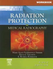
Covers radiation biology, dose limits, and regulatory requirements in accessible terms. Positions you to meet the radiation-safety and licensure knowledge requirements before moving to board prep.
Clinical Procedures & Patient Care
IntermediateMaster the full scope of nuclear medicine clinical procedures — patient preparation, injection techniques, imaging protocols, and artifact recognition — as performed by a practicing technologist.
▸ Study plan for this stage
Pace: 8–10 weeks, ~40–50 pages/day, with 2–3 dedicated lab/clinical observation days per week
- Patient preparation protocols and safety considerations for nuclear medicine procedures (fasting, medication hold times, pregnancy screening, radiation safety)
- Radiopharmaceutical selection, handling, quality control, and dose calculation specific to different organ systems and clinical indications
- Venipuncture and injection techniques: proper IV access, bolus vs. infusion administration, extravasation recognition and management
- Imaging acquisition protocols for major procedures (myocardial perfusion, bone scan, renal imaging, thyroid uptake, gastric emptying, etc.) including positioning, timing, and camera settings
- Artifact recognition and troubleshooting: motion artifacts, attenuation, scatter, collimator artifacts, and technical errors that compromise image quality
- Quality assurance and quality control procedures for gamma cameras, dose calibrators, and radiopharmaceutical preparations
- Adverse reaction recognition and emergency response protocols specific to radiopharmaceutical administration
- Documentation, communication with physicians, and integration of nuclear medicine findings into clinical decision-making
- What are the essential pre-procedure patient preparation steps for a myocardial perfusion study, and why is each step clinically important?
- How do you select and prepare the appropriate radiopharmaceutical for a given clinical indication, and what quality control checks must be performed before administration?
- Describe the proper venipuncture technique and injection procedure for nuclear medicine, including how to recognize and manage extravasation.
- What are the key imaging acquisition parameters (timing, positioning, energy windows, collimator selection) for at least three major procedures covered in the text?
- How do you identify common artifacts in nuclear medicine images, determine their cause, and implement corrective measures?
- What are the critical quality assurance procedures for gamma camera systems, and how do you interpret QA results to ensure diagnostic image quality?
- Create a detailed patient preparation checklist for three different nuclear medicine procedures (e.g., myocardial perfusion, bone scan, thyroid uptake), including timing, medications to hold, and safety screening questions.
- Practice radiopharmaceutical dose calculations using realistic clinical scenarios; verify calculations against a dose calibrator protocol or reference table.
- Observe or simulate venipuncture and IV injection technique with a phantom arm or under clinical supervision; document proper technique and potential complications.
- Review and analyze 10–15 clinical images from Ahmadzadehfar's text or institutional archives; identify at least two artifacts per image, classify them (motion, attenuation, scatter, etc.), and propose corrective measures.
- Perform a complete gamma camera quality control protocol (uniformity, spatial resolution, energy resolution, linearity) and interpret results against acceptance criteria; document findings.
- Conduct a mock clinical case presentation: given a patient history and clinical question, select the appropriate procedure, outline the protocol, predict expected findings, and discuss potential artifacts or pitfalls.
Next up: This stage equips you with hands-on mastery of standard clinical procedures and real-world troubleshooting skills, preparing you to advance to specialized imaging modalities, advanced protocols (SPECT/CT, PET), and complex case interpretation in the next stage.

A case-rich, clinically detailed text that reinforces how imaging findings correlate with disease states, sharpening image-interpretation skills needed for both practice and the boards.
Discussion
Keep reading
Paths that share books, cover the same subject, or open a related topic.