Radiology and Medical Imaging: The Best Books to Read, in Order
Radiology is one of the few medical specialties you can learn a great deal about from books alone, because the whole discipline is about pattern recognition on a flat image and the standard teaching texts are built to train exactly that. This path runs from the introductory texts a first-year medical student is handed, through the chest film (still the single most-ordered study in medicine, and the one every clinician is expected to read), out to the cross-sectional reference works and finally to the physics that explains why an X-ray, a CT and an MRI show completely different things. Two honest warnings before you start. These are clinical textbooks, not popular science: expect anatomy vocabulary from page one, and expect to buy expensively or borrow from a library. And several of the records here are catalogued at an older edition than the one currently in print, which is noted book by book below — for the introductory texts the edition barely matters, but for CT, MRI and anything touching dose or protocol you want the newest edition you can get.
The Introductory Texts
BeginnerLearn what each imaging modality actually measures, how a radiograph is produced and described, and the systematic search pattern that stops you seeing only the obvious abnormality. The stage opens with a general-audience history and then moves to texts written at medical-student level, which assume basic gross anatomy but nothing about imaging; work through them before anything below. Note that the catalogue records for several of these sit at earlier editions than the current print run, which
▸ Study plan for this stage
Pace: Six to eight weeks, and the four books do not read at the same rate. Kevles's Naked to the Bone is 378 pages of general-audience narrative history and will go at 40-50 pages an evening; read it first and finish it in a fortnight. The three teaching texts cannot be read that way. Radiology 101 has no
- What each modality physically measures: X-ray attenuation, acoustic reflection, tissue proton behaviour in a magnetic field, radiotracer uptake - and why the same organ looks different in each
- The vocabulary that follows from that: radiodense and radiolucent for plain film and CT, hypo- and hyperechoic for ultrasound, hypo- and hyperintense for MRI, and why the terms are not interchangeable
- A fixed systematic search pattern, applied in the same order every time, as the thing that stops you stopping at the first abnormality
- How a radiograph is actually produced - beam, patient, detector - and why projection, penetration and patient position change what appears on the film
- Describing a finding before naming it: location, density, margin, size, effect on surrounding structures
- Which study answers which clinical question, and the ordering decision as a real skill separate from interpretation
- The history in Kevles as context for a live constraint: each modality arrived with its own dose, cost and consent problem, and those problems are still what limits its use
- Given a lesion that is bright on CT and dark on a T2-weighted MRI, what does each observation tell you about the tissue, and why do the two answers not contradict each other?
- What is the search pattern you now use on a plain film, and what does each step in it exist to catch?
- Erkonen and Smith teach the modalities one at a time before showing a case. What does that ordering give you that a case-led book cannot?
- Herring builds each chapter as one pattern shown normal and then broken. Pick a pattern you have learned that way and state what the normal appearance is that makes the abnormal one visible.
- Which of the studies covered here involve ionising radiation, and how would that change which one you would order first in a young patient?
- Take one image of each modality from Radiology 101 and write a single sentence for each saying what physical property produced the contrast you are looking at. Then check your sentence against Erkonen and Smith's own explanation and note where you reached for the wrong mechanism.
- Work through Herring in his order with the caption covered. Describe the finding aloud - location, density, margin - before you uncover the answer. Herring's normal-then-broken structure only teaches recognition if you do not read the answer first.
- Read Singh's Radiology Fundamentals over the same body systems you have just finished in Herring, and keep a list of everything Singh says that Herring does not. Two passes over one territory in different voices is the reason both books are on this stage.
- Write out your search pattern on an index card and use it on every image in the last chapter of Herring you read. Count how many times the pattern found something after you had already spotted the obvious abnormality.
- Follow one modality through Kevles from invention to routine use - CT is the clearest thread - then find where that modality's characteristic artefacts appear in Herring or Singh. The physical compromise made in the design is usually the artefact you are being taught to recognise.
Next up: With a search pattern and the vocabulary of all five modalities in place, the next stage narrows to the single study every clinician is expected to read unaided and drills it until the routine is automatic.
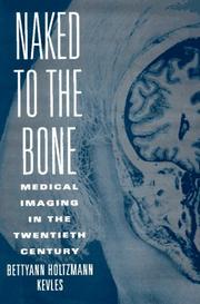
The one narrative history on this path and the right way in: how X-rays, ultrasound, CT, MRI and PET were invented and how each changed what medicine could see and what patients would consent to. No technical background needed, and it gives the textbooks that follow a reason to exist. Note the record resolves under the author's name without her middle name.
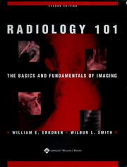
The gentlest possible entry: it teaches the modalities one at a time — plain film, ultrasound, CT, MRI, nuclear medicine — and explains what physical property each one is measuring before it shows you a single case. Start here if the difference between a hypodense and a hypointense lesion is not yet second nature.

The book most students actually learn from, and the best single purchase on this path. Herring teaches recognition rather than lists: each chapter builds one pattern, shows it normal, then shows it broken. Catalogued here under the bare title; the same work is published as Learning Radiology: Recognizing the Basics, so buy whichever edition is current rather than both.

A short, deliberately non-exhaustive companion aimed at students and non-radiologist clinicians who need to order and interpret studies competently. Read it alongside Herring as a second pass over the same ground in a different voice.
Reading the Chest Film
IntermediateBecome genuinely competent at the one study every doctor is expected to read unaided. By the end you should have a fixed search order for a chest radiograph, be able to localise a lung opacity by the silhouette sign, and know the handful of findings on a trauma film that change management in the next ten minutes. This stage assumes the first one; the emergency book in particular is written for people already comfortable with normal anatomy.
▸ Study plan for this stage
Pace: Eight to ten weeks for 1,121 pages, and this is the stage where reading rate is the wrong measure. Corne's Chest X-Ray Made Easy is only 156 pages and can be finished in a week - it exists to hand you a search order, and it is deliberately placed first for that reason. De Lacey, Morley and Berman's
- A fixed search order for the chest film, run identically every time, ending with the review areas that are habitually skipped - apices, behind the heart, below the diaphragm, the bones
- The silhouette sign: loss of a normal interface localises an opacity to the lobe abutting it, which is what lets you say where rather than merely that
- The air bronchogram as the marker of airspace rather than interstitial or pleural disease
- Normal appearances that mimic pathology - vessels, nipples, skin folds, companion shadows - as the main source of false positives
- Assessing film adequacy first: rotation, inspiration, penetration, and whether the study is supine or erect, because each changes what is visible
- The handful of trauma findings that change management immediately, and the discipline of the second look that Raby's book is built around
- Reading the film against the clinical question rather than in the abstract
- An opacity obscures the right heart border but the right hemidiaphragm is sharp. Where is it, and what is the reasoning?
- How do you distinguish an airspace process from an interstitial one on a plain film, and which single sign does most of that work?
- What are the review areas in your search pattern, and what is the commonest miss in each?
- A supine trauma film in a patient with a pneumothorax may look normal at the apex. Why, and where do you look instead?
- Felson is written as a programmed text rather than a reference. What does the question-and-answer format teach that a chapter of prose on the same material would not?
- Raby's book is organised around commonly missed injuries rather than by anatomy. What does that ordering assume about the reader?
- Write Corne's search routine on a card and apply it, in that exact order, to every case in de Lacey, Morley and Berman before you read their commentary. Log each case as hit, miss or false positive, and at the end read back the misses as a group - they will cluster in one or two review areas.
- Work Felson with the answers physically covered, and write your answer down before uncovering. A programmed text scores you honestly only if the answer is committed first; the format is the exercise.
- Use the silhouette sign as Felson develops it to localise every lung opacity in de Lacey, Morley and Berman, stating the lobe before checking. This is the one drill that converts Felson's reasoning into Corne's routine.
- Give yourself thirty seconds on each of ten cases from Raby, De Lacey and Berman, then a full second look with your search pattern. The gap between the two passes is the argument for the pattern, measured on yourself.
- Collect the normal mimics shown in de Lacey and Corne - skin folds, companion shadows, vessels seen end-on - into one list, then go back through the Raby cases specifically hunting for them among your false positives.
Next up: Having mastered the projection image, the next stage moves off the plain film entirely - to the whole body, to cross-sectional anatomy, and to reference texts used by system and by problem rather than read through.

The shortest useful book on the chest film and the right first pass: a systematic routine, then the common patterns, in about a hundred and fifty pages.
De Lacey, Morley and Berman's survival guide: a large-format, case-led book organised around the findings that are actually missed, with the normal appearances that mimic them shown alongside. The practical drill between Corne's routine and Felson's reasoning.
The classic programmed text on the chest film, built around the silhouette sign and the air bronchogram — the two ideas that let you say where in the chest an opacity sits rather than merely that it is there. It is taught in a question-and-answer format that rewards working through it slowly rather than reading it. The record here is an earlier edition; the book has been revised several times and the current one is worth seeking.
The natural application of everything above, and the book that teaches the discipline of the second look: it is organised around the injuries that are commonly missed on a first pass. Read it after Felson, because it assumes you already have a search pattern to apply.
The Reference Texts and Cross-Sectional Imaging
IntermediateMove from the plain film to the whole body and to CT and MRI, and learn to use a reference text the way a working radiologist does — by system and by problem, not cover to cover. By the end you should be able to orient yourself in an axial CT of the abdomen and explain why a particular MRI sequence was chosen. These are large, expensive books; a library or a departmental copy is the sane approach, and edition currency matters much more here than in stage one, because protocols and dose guidance
▸ Study plan for this stage
Pace: Three to four months, and the way you handle these 3,055 pages matters more than the pace. Novelline's Squire's Fundamentals of Radiology is 638 pages and is the only one of the four still readable straight through - do that first, a chapter a sitting, over about six weeks, to get a map of the terri
- Cross-sectional orientation: reading an axial stack as a volume, knowing which way is left, and following a structure through consecutive slices rather than reading one image
- CT attenuation in Hounsfield units, and windowing as the decision about which range of that scale you are looking at - the same slice is a lung, soft tissue or bone image depending on it
- What intravenous and enteric contrast add, what phase of enhancement a study was taken in, and why the phase determines what a lesion looks like
- T1 and T2 weighting, and the small set of sequences that answer most clinical questions, with fat suppression and diffusion as the common problem-solvers
- Choosing between CT and MRI on the basis of what each measures, plus speed, availability, dose and contrast risk
- Using a reference text by system and by problem - Brant and Helms as lookup, not as reading
- Reading a study against its protocol: what was requested, what was acquired, and what the acquisition could not have shown
- Why does the same abdominal CT slice look completely different in soft-tissue and lung windows, and what is being discarded in each?
- A lesion is bright on T1 without contrast. What are the candidate explanations, and which additional sequence would separate them?
- Webb and Brant teach CT anatomy plane by plane before any pathology. What goes wrong for a reader who inverts that order?
- For a given clinical question - a suspected renal mass, say - would you choose CT or MRI, and what specifically does the losing modality fail to give you?
- Novelline is organised by body system and Brant and Helms is organised as a reference. Which one do you reach for when you have a finding and no diagnosis, and why?
- Take one body system - the abdomen is the most useful - and read Novelline's chapter, then Brant and Helms's coverage of the same system, then the corresponding chapters of Webb and Brant. Write down what each of the three adds that the others do not. That comparison is the whole argument for having all three.
- Work through Webb and Brant's normal-anatomy chapters by naming every labelled structure on a slice with the labels covered, moving up and down the stack until you can follow a single vessel through consecutive images.
- For ten cases in Brant and Helms, note the imaging protocol described - modality, contrast, phase - and write one sentence saying what that protocol was chosen to demonstrate and what it would have missed.
- Work the sequence-selection material in Westbrook and Talbot against real problems: for four clinical questions, state which sequences you would want and what each contributes. Then check your answer against the book's own account of what each sequence weights.
- Use Brant and Helms deliberately as lookup rather than reading - take twenty findings you met in stage two's chest films and find each one in the reference, timing yourself. Fluency with the index is a real skill and it only comes from use.
Next up: Every trade-off met so far - resolution against dose, contrast against noise, speed against signal - has been stated as a rule; the final stage replaces the rules with the physics that produces them.
The bridge from the introductory texts to the reference ones: broader than Herring, organised by body system, and still readable straight through. Take it before the larger works so you have a map of the territory they cover. The catalogued record is an older edition of a long-running text.
Brant and Helms is the standard single-source reference for residents and the book most departments keep on the shelf — comprehensive by system, with enough explanation to learn from rather than merely look things up. This is the reference text of the path; the record is a mid-2000s edition, and later ones expand the cross-sectional and MRI material considerably.
The focused companion to Brant for the modality that now carries most of the diagnostic load. Webb teaches CT anatomy plane by plane before pathology, which is the correct order and the one most self-taught readers skip.

MRI is the one modality you cannot use well without understanding how the image is produced, and this is the standard text that explains T1, T2, sequence selection and artefact in terms a clinician can follow. It sits here rather than in the physics stage because it is written for practitioners, and it is the natural handover into that stage.
How the Machines Actually Work
IntermediateUnderstand the physics well enough to reason about contrast, resolution, artefact and dose rather than memorising rules. This stage is genuinely harder than everything above and assumes comfort with basic physics and maths; it is optional for a clinician and compulsory for anyone going into the specialty or into medical physics. Edition age is a real problem in this stage specifically — the underlying physics does not change, but equipment, dose figures and regulatory guidance do, so treat any d
▸ Study plan for this stage
Pace: Four to six months for 1,320 pages, and this is by a wide margin the hardest stage on the path. Curry's Christensen's Physics of Diagnostic Radiology is 522 pages and comes first because it is the more forgiving of the two: it is the standard teaching text for radiology trainees and explains image q
- X-ray production: the tube, the bremsstrahlung and characteristic spectrum, and how kVp and mAs each change the beam in different ways
- Beam-matter interaction - photoelectric absorption and Compton scatter - as the origin of subject contrast and of the scatter that destroys it
- The image-quality triangle: contrast, noise and spatial resolution as quantities traded against each other and against dose, never optimised independently
- Dose quantities and where they differ: absorbed, equivalent and effective dose, and why the same exposure means different things for different tissues
- Digital detection and the fact that a digital image cannot be judged over- or underexposed by its appearance, which is the modern dose problem
- CT reconstruction from projections, and what pitch, collimation and reconstruction algorithm each do to the resulting image
- The physical origin of the common artefacts, so that an artefact is diagnosed from its mechanism rather than matched to a picture
- Where MRI physics departs from everything else: no ionising radiation, contrast generated by relaxation behaviour rather than attenuation, and an entirely different safety problem
- Raising kVp and raising mAs both brighten the image. Why are they not interchangeable, and what happens to contrast and to dose with each?
- Why is scatter the dominant limit on radiographic contrast in a thick body part, and what do a grid, collimation and an air gap each do about it?
- Curry teaches image quality as a set of trade-offs. State one trade-off in your own terms and say what you would give up to improve resolution in a specific study.
- Johns treats dosimetry in far more depth than Curry. What does absorbed dose fail to tell you that equivalent and effective dose are introduced to capture?
- Pick one CT artefact and explain it from the physics up rather than by appearance. What acquisition change would reduce it, and what would that cost?
- Both of these records are old printings. Which parts of each remain trustworthy, and which specifically must be replaced from a current source?
- Work the derivations in Curry with a pen rather than reading past them - attenuation, exponential absorption, the relationship between kVp and beam quality. This stage's difficulty is entirely in the derivations, and skimming them produces a reader who can quote the trade-offs but cannot use them.
- Reproduce one of Curry's own worked calculations with its own numbers, then re-run it with a single parameter changed and predict the effect on contrast, noise and dose before checking against the text.
- For each of the artefacts covered in Curry, write a two-line entry giving the physical mechanism and the acquisition change that reduces it, then find the same artefact in the cases of the previous stage's cross-sectional books and confirm your mechanism explains what you see.
- Take the dose figures quoted in Curry and Johns and compare each against a current published reference for the same study. The exercise is not the number but the direction and size of the drift - it is the clearest demonstration of why edition currency matters in this stage and not the others.
- Read Johns's treatment of radiation interaction alongside Curry's on the same topic and write down what the extra rigour buys. Where Curry gives a rule, Johns usually gives the quantity the rule approximates; naming three such pairs is the point of reading both.
Next up: This is the end of the path: with the physics in place, the earlier stages' rules become consequences you can re-derive, and further progress comes from reading current editions and reported studies rather than more textbooks.
The standard teaching text on imaging physics for radiology trainees, and the one that explains image quality — contrast, noise, resolution, scatter — as a set of trade-offs rather than a list of facts. Read it before Johns; it is the more forgiving of the two. The record is a 1990 edition, so use it for the physics and not for equipment specifics.
The classic and more rigorous treatment, covering radiation production, interaction and dosimetry in far more depth, and reaching into radiotherapy physics as well. Flagged plainly: the catalogued record here is a very early edition of a book that ran for decades, so read it as a foundational text and take anything equipment- or dose-specific from a current source.
Discussion
Keep reading
Paths that share books, cover the same subject, or open a related topic.