MRI and CT technologist: books to break into medical imaging
This curriculum builds from zero medical knowledge to ARRT registry-ready competency across four progressive stages. It begins with the language of anatomy and medical imaging, advances through the physics and protocols of both MRI and CT, and culminates in targeted registry review — mirroring the knowledge arc of an accredited radiologic technology program.
Foundations: Anatomy & Medical Language
BeginnerBuild the anatomical vocabulary and body-system knowledge that every imaging concept will depend on — so that terms like 'axial slice through L4' or 'coronal view of the mediastinum' are immediately meaningful.
▸ Study plan for this stage
Pace: 8–10 weeks, ~40–50 pages/day (alternating between books: 2–3 weeks on "The Language of Medicine" for terminology foundation, then 5–7 weeks on "Gray's Anatomy for Students" for detailed anatomical systems)
- Medical terminology: word roots, prefixes, suffixes, and how they combine to describe anatomy and pathology (e.g., 'gastro-' + '-itis' = inflammation of the stomach)
- Anatomical planes and directional terms (axial, coronal, sagittal; superior, inferior, medial, lateral, anterior, posterior) as the foundation for interpreting cross-sectional imaging
- Body cavities and regions: thoracic, abdominal, pelvic, and how organs are organized within them
- Major organ systems and their components: skeletal, muscular, nervous, cardiovascular, respiratory, digestive, urinary, endocrine, reproductive, and lymphatic
- Gross anatomy of the trunk, head, neck, and limbs with emphasis on surface landmarks and bony reference points used in imaging protocols
- Organ relationships and spatial anatomy: how structures relate to each other in three dimensions (e.g., the pancreas posterior to the stomach, the spinal cord within the vertebral canal)
- Normal anatomical variants and common anatomical relationships that appear in imaging slices
- Imaging-relevant anatomy: understanding how 2D cross-sections relate to 3D anatomy, and why certain structures appear in specific imaging planes
- What do the prefixes 'hyper-', 'hypo-', 'tachy-', and 'brady-' mean, and how do they modify medical terms? Provide 3–4 examples relevant to imaging findings.
- Define the anatomical planes (axial, coronal, sagittal) and directional terms (medial, lateral, superior, inferior, anterior, posterior). Explain how each plane cuts through the body and what structures you would see in each.
- Name the major organs in the thoracic cavity and describe their relationships to each other. Which organs are anterior, posterior, medial, and lateral?
- Describe the layers of the abdominal wall and the peritoneal cavity. What is the difference between intraperitoneal and retroperitoneal organs, and why does this matter in imaging?
- Trace the path of the esophagus, stomach, small intestine, and colon through the abdomen and pelvis. At what vertebral levels do key transitions occur?
- Identify the major arteries and veins of the trunk (aorta, IVC, portal vein, etc.) and describe their courses. Why is vascular anatomy critical for interpreting CT and MRI?
- Terminology building: Create flashcards for 50–75 medical word roots, prefixes, and suffixes from 'The Language of Medicine.' Group them by body system and quiz yourself daily until you can instantly recognize them in clinical terms.
- Anatomical plane practice: Using Gray's Anatomy diagrams, select 10 anatomical structures (e.g., liver, pancreas, kidneys, heart, lungs) and sketch or describe what you would see in axial, coronal, and sagittal planes at different levels.
- 3D visualization: For each major organ system, use Gray's Anatomy illustrations to mentally 'walk through' the anatomy in three dimensions. Then close the book and verbally describe the spatial relationships without looking.
- Labeling exercises: Print or download unlabeled anatomical diagrams from Gray's Anatomy (or use online anatomy atlases) and label structures in axial, coronal, and sagittal views. Repeat for thorax, abdomen, pelvis, and head/neck.
- Clinical correlation: Read 5–10 simple imaging case descriptions (e.g., 'axial CT through L4 shows the pancreas anterior to the left kidney'). For each, sketch or describe what you expect to see and verify against Gray's Anatomy illustrations.
- Vertebral level mapping: Create a reference chart showing what organs and structures appear at key vertebral levels (T1, T4, T6, T10, L1, L3, L5, S1). Use Gray's Anatomy cross-sectional images to build this chart.
Next up: Mastery of anatomical terminology and 3D spatial relationships prepares you to understand imaging protocols and recognize normal anatomy in actual MRI and CT scans—the foundation for identifying pathology in the next stage.
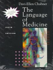
The single best-selling medical terminology text for allied health students; reading it first gives you the prefix/suffix/root system that unlocks every clinical term you will encounter in imaging.
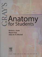
A clinically oriented, richly illustrated anatomy reference that introduces regional and cross-sectional relationships — the spatial thinking that CT and MRI interpretation demands.
Physics & Technology: How the Machines Work
IntermediateUnderstand the physical principles behind CT (X-ray attenuation, Hounsfield units, reconstruction) and MRI (spin physics, pulse sequences, k-space) well enough to make protocol decisions and troubleshoot image quality.
▸ Study plan for this stage
Pace: 8–10 weeks, ~25–30 pages/day, with 2–3 days per week dedicated to concept review and practical application
- Nuclear magnetism and the behavior of hydrogen nuclei in a magnetic field (precession, Larmor frequency, resonance)
- Longitudinal (T1) and transverse (T2) relaxation times and their tissue-dependent values
- Radiofrequency (RF) pulses and their role in tipping magnetization (flip angles, 90° and 180° pulses)
- Pulse sequences as the foundation of image contrast (spin echo, gradient echo, inversion recovery, fast sequences)
- K-space and its relationship to spatial frequency, phase encoding, and frequency encoding
- Reconstruction from k-space data and how it determines spatial resolution and image artifacts
- Signal-to-noise ratio (SNR) and contrast-to-noise ratio (CNR) as drivers of protocol optimization
- Common artifacts (motion, aliasing, susceptibility, chemical shift) and their physical origins
- Explain how hydrogen nuclei behave in a magnetic field and why the Larmor frequency is critical to MRI physics
- Describe the difference between T1 and T2 relaxation and how tissue-specific relaxation times create image contrast
- How do 90° and 180° RF pulses manipulate magnetization, and what is the purpose of each in a spin echo sequence?
- What is k-space, how is it filled during a pulse sequence, and how does the trajectory through k-space affect image quality and acquisition time?
- Given a clinical scenario (e.g., suspected stroke, knee ligament tear), explain which pulse sequence you would choose and why, based on tissue contrast and SNR requirements
- Identify the physical cause of a specific artifact (e.g., motion blur, aliasing, susceptibility artifact) in an MR image and propose a protocol adjustment to mitigate it
- Create a comparison table of at least 5 pulse sequences (spin echo, fast spin echo, gradient echo, FLAIR, STIR) showing TR, TE, flip angle, and the tissue contrast each produces; annotate with clinical use cases
- Work through 3–5 k-space diagrams: sketch how phase and frequency encoding fill k-space, then predict how changing matrix size, FOV, and phase encoding steps affects resolution and acquisition time
- Analyze 5–10 clinical MR images (from textbook or online repository) and identify the pulse sequence used, the tissue contrast visible, and any artifacts present; justify your reasoning
- Solve 10–15 quantitative problems on Larmor frequency, relaxation time calculations, SNR, and flip angle effects using realistic field strengths (1.5T, 3T) and tissue parameters
- Design a complete MRI protocol for 3 different clinical scenarios (e.g., brain stroke, knee meniscus tear, liver lesion) specifying pulse sequences, TR/TE values, flip angles, and field-of-view; justify each choice
- Troubleshoot 5 image quality problems: given a degraded image and clinical context, identify the likely cause (physics-based) and propose a protocol modification
Next up: Mastery of MRI physics and pulse sequences equips you to understand advanced imaging techniques (parallel imaging, diffusion, perfusion, spectroscopy) and to recognize how hardware limitations and safety constraints shape protocol design in the next stage.

The most widely used MRI physics and protocols book worldwide; its step-by-step explanations of spin, relaxation, pulse sequences, and artifacts are accessible to newcomers yet thorough enough for registry prep.
Registry Prep & Clinical Integration
ExpertConsolidate all prior knowledge into exam-ready, clinically applicable competency — passing the ARRT Advanced CT and/or MRI registry examinations and performing confidently in the clinical environment.
▸ Study plan for this stage
Pace: 8–10 weeks, ~40–50 pages/day (with active review and practice exams interspersed)
- CT physics fundamentals: X-ray production, attenuation, beam geometry, and image reconstruction algorithms (filtered back-projection, iterative reconstruction)
- Hounsfield units and CT number calculation: how tissue density relates to pixel values and clinical interpretation
- Detector technology and scanner geometry: evolution from single-slice to multi-detector CT (MDCT), helical scanning, and data acquisition modes
- Image quality parameters: spatial resolution, contrast resolution, noise, artifacts, and dose optimization strategies
- Radiation dose in CT: CTDI, DLP, effective dose, dose reduction techniques, and regulatory compliance (ACR, ASRT standards)
- Clinical protocols and scanning techniques: patient positioning, contrast administration timing, bolus tracking, and organ-specific acquisition parameters
- Artifact recognition and mitigation: metal, motion, beam hardening, and ring artifacts—causes and clinical solutions
- Quality assurance and safety: daily/weekly/monthly QA procedures, equipment maintenance, and infection control in the CT environment
- Explain how filtered back-projection and iterative reconstruction differ in image formation, and describe when each is clinically preferred.
- Calculate or interpret Hounsfield units for common tissues (bone, soft tissue, fat, air) and explain how HU values affect diagnostic image quality.
- Describe the advantages of multi-detector CT (MDCT) over single-slice CT in terms of speed, coverage, and dose efficiency.
- Identify common CT artifacts (metal, motion, beam hardening, ring), explain their causes, and propose practical mitigation strategies.
- Design a CT protocol for a specific clinical scenario (e.g., abdominal trauma, pulmonary embolism) including patient prep, scanning parameters, contrast timing, and dose justification.
- Interpret CTDI and DLP values, calculate effective dose, and explain how to optimize scanning parameters to reduce patient radiation exposure while maintaining diagnostic quality.
- Complete end-of-chapter review questions and case studies in Romans' textbook; track weak areas for targeted re-review.
- Perform hands-on QA testing on your institution's CT scanner (CTDI phantom scans, spatial resolution assessment, noise measurement) and document results against baseline standards.
- Analyze 10–15 clinical CT images from your institution: identify artifacts, assess image quality, and propose protocol adjustments to improve future scans.
- Simulate registry exam conditions: take full-length ARRT CT practice exams under timed conditions; review incorrect answers against Romans' chapters and ARRT content specifications.
- Create protocol cards or a digital reference guide for 5–8 high-frequency clinical scenarios (chest, abdomen, pelvis, neuro, trauma) with specific parameters, contrast protocols, and dose estimates.
- Conduct peer teaching: explain CT physics concepts (reconstruction, artifacts, dose) to a colleague or student; refine explanations based on their questions and feedback.
Next up: Mastery of CT physics, clinical protocols, and quality assurance positions you to pass the ARRT Advanced CT registry exam and perform as a confident, independent CT technologist—and provides a foundation for advanced cross-sectional imaging roles (e.g., MRI technologist, interventional technologist, or imaging supervisor) in subsequent career stages.

Bridges physics and clinical practice by covering positioning, protocols, pathology recognition, and patient care — a practical capstone that ties together everything learned and prepares you for day-one clinical competency.
Discussion
Keep reading
Paths that share books, cover the same subject, or open a related topic.